Benefits of Having an Ultrasound at a Local Facility

When your doctor recommends diagnostic imaging, convenience probably matters more than you initially realise. Between work, school runs, family responsibilities, Johannesburg traffic, and everything else competing for your time, travelling across the city for a medical appointment can turn a relatively straightforward examination into a much bigger undertaking.

That is why so many patients begin by searching for an ultrasound near me

Choosing a local ultrasound facility is not simply about saving a few kilometres. It can make it easier to attend your appointment, reduce unnecessary travel, simplify follow-up care, and provide access to professional diagnostic imaging within a familiar healthcare environment.

Diagnostic Radiological Services (DRS) provides ultrasound and a broad range of diagnostic imaging services across convenient locations in Johannesburg’s eastern and southern areas. We aim to make the imaging process as efficient, comfortable, and straightforward as possible while providing the diagnostic information your referring healthcare professional needs.

What Is an Ultrasound?

Ultrasound, sometimes called sonar, is a medical imaging technique that uses high-frequency sound waves to create images of structures inside the body.

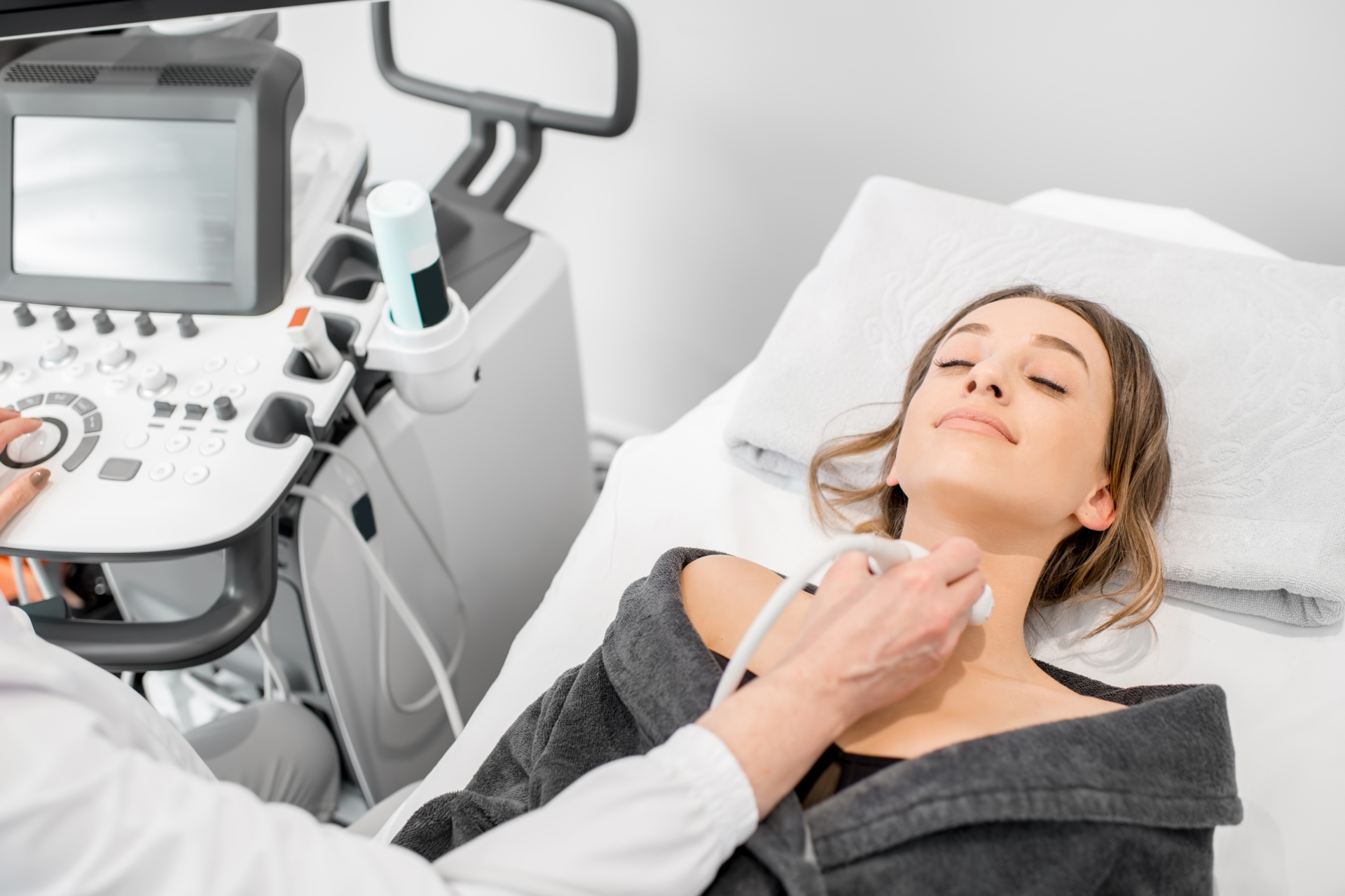
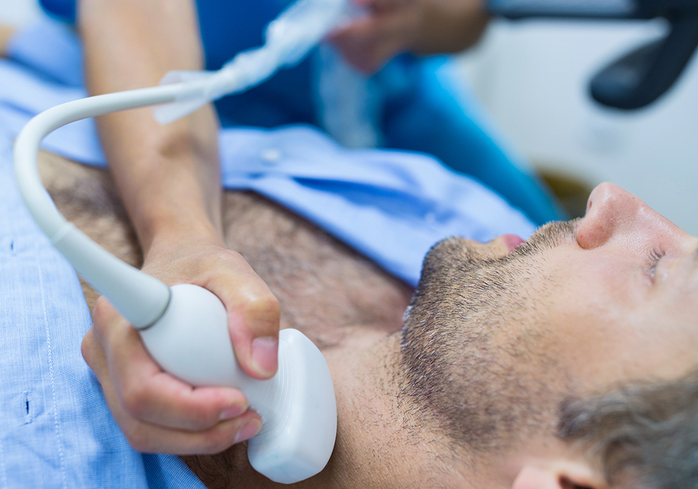
A small handheld device called a transducer sends sound waves into the area being examined. These waves are reflected differently by various tissues and structures, allowing the ultrasound equipment to create images that can be viewed in real time.

Unlike X-rays and CT scans, ultrasound does not use ionising radiation.

This makes it an extremely useful imaging method for examining many parts of the body and investigating a wide variety of medical concerns.

A specialised form of ultrasound called Doppler ultrasound can also be used to assess blood flow through blood vessels.

Your doctor will determine whether an ultrasound is the most appropriate imaging examination based on your symptoms, medical history, and the clinical information they need.

Why Search for an Ultrasound Near Me?

When you type ultrasound near me  into a search engine, you are probably looking for more than the closest facility on a map.

You want somewhere that is convenient, professional, easy to access, and equipped to provide the diagnostic imaging your doctor has requested.

There are several practical advantages to choosing a local facility.

1. You Spend Less Time Travelling

Anyone who regularly drives around Johannesburg or the East Rand knows that a seemingly short journey can quickly become a lengthy one when traffic joins the party.

Choosing a local ultrasound facility means potentially spending less time travelling to and from your appointment.

This can be particularly helpful if you need to schedule your ultrasound around work, collect children from school, depend on somebody else for transport, or simply do not feel well enough for a long journey.

When healthcare is easier to access, attending necessary appointments becomes easier too.

2. It Is Easier to Fit Your Ultrasound Into a Busy Day

Medical appointments rarely happen on days when you have nothing else planned.

For many South Africans, an ultrasound appointment needs to fit somewhere between meetings, family responsibilities, errands, and commuting.

Having a facility close to home or work can make the process considerably more manageable. Instead of setting aside a large portion of your day for travel, you can focus on attending the examination itself and getting back to your routine.

That convenience may seem like a small detail, but when you are already worried about your health, fewer logistical headaches can make a meaningful difference.

3. Local Diagnostic Imaging Can Feel Less Intimidating

It is completely normal to feel a little apprehensive before a medical examination, particularly when you are waiting for answers about an unexplained symptom or health concern.

Being treated somewhere familiar can help.

You may already know the hospital, neighbourhood, or surrounding area, which means there is one less unknown to deal with on the day.

We aim to create a professional, patient-focused experience from the moment you arrive. Our teams understand that diagnostic imaging is routine for us, but it may be unfamiliar or worrying for you.

We want you to know what to expect and feel comfortable asking questions about the examination process.

4. Ultrasound Does Not Use Ionising Radiation

One of the most important benefits of ultrasound is that it creates images using sound waves rather than ionising radiation.

This allows ultrasound to provide valuable real-time information about many soft tissues, organs, structures, and blood vessels.

It is one reason ultrasound is used so widely throughout modern medicine.

However, ultrasound is not automatically the best examination for every medical problem. Depending on what your doctor needs to investigate, another imaging modality, such as an X-ray, CT scan, MRI, mammogram, or bone density examination, may be more appropriate.

Diagnostic Radiological Services offers a comprehensive range of diagnostic radiology services, allowing different imaging technologies to be used according to the clinical question that needs to be answered.

5. Ultrasound Can Provide Real-Time Imaging

Ultrasound differs from many other imaging examinations because images can be viewed as the examination is taking place.

This real-time capability makes ultrasound particularly useful for assessing moving structures and evaluating blood flow when Doppler techniques are used.

The precise examination performed will depend on the referral from your healthcare professional.

Rather than approaching an ultrasound as a generic scan, your imaging team works from the clinical question provided by your doctor. This helps ensure that attention is directed towards the area and information relevant to your care.

6. A Local Facility Can Make Follow-Up Care Simpler

Diagnostic imaging is often only one part of a patient’s healthcare journey.

After the ultrasound, the images need to be assessed and the findings communicated so that your referring healthcare professional can consider them alongside your symptoms, medical history, examination, and any other test results.

A radiologist formulates a report following the relevant imaging examination. Your results are then made available through our practice or your referring doctor, depending on the process involved.

Your referring healthcare professional can discuss the findings with you in the context of your overall health and advise you about what happens next.

Having your imaging performed locally may make the entire journey easier, particularly if additional appointments or investigations are required.

7. You Have Access to a Broader Range of Diagnostic Imaging Services

Sometimes an ultrasound answers the clinical question. At other times, additional imaging may be recommended.

This is one of the advantages of choosing an established diagnostic radiology practice rather than viewing each scan as a completely separate healthcare event.

Diagnostic Radiological Services provides access to a range of diagnostic imaging services, which include ultrasound as well as X-ray, mammography, bone densitometry, MRI, CT, PET-CT, cone beam imaging, and interventional radiology services at selected facilities.

Not every imaging modality is available at every branch, so it is always worth checking which Diagnostic Radiological Services location provides the examination you require.

Our network gives patients across Johannesburg’s east and south access to professional diagnostic imaging at conveniently situated facilities.

What Can an Ultrasound Be Used For?

Ultrasound is a versatile diagnostic imaging technique.

Doctors may request ultrasound imaging to investigate symptoms or assess structures and organs in different parts of the body. Depending on the clinical situation, ultrasound may be useful when examining soft tissues, abdominal or pelvic structures, blood vessels, or other areas that can be effectively visualised using sound waves.

It is also widely known for its role in pregnancy imaging.

Doppler ultrasound provides additional information by helping assess the movement of blood through vessels.

The important point is that the examination should be matched to the medical question being investigated.

If your doctor has requested an ultrasound, the referral provides the imaging team with important information about the examination required and why it is being performed.

What Should You Expect During an Ultrasound?

The exact process depends on the ultrasound your doctor has requested.

For many examinations, you will be positioned so that the area being investigated can be easily accessed. A gel is generally applied to the skin to help the ultrasound transducer make good contact with the body and transmit sound waves effectively.

The transducer is then moved across the relevant area while images are produced on a monitor.

You may be asked to change position, remain still, or follow simple instructions during the examination so that the necessary images can be obtained.

Some pressure may be required when scanning certain areas. If the area is already tender, this may feel temporarily uncomfortable.

Your imaging professional will guide you through the procedure.

Do I Need to Prepare for an Ultrasound?

Possibly.

Preparation requirements vary depending on the type of ultrasound being performed. Some examinations require particular preparation to make certain organs or structures easier to visualise, while others require very little preparation.

For that reason, do not assume that all ultrasound appointments work the same way.

When arranging your Diagnostic Radiological Services appointment, make sure you understand the preparation instructions for your particular examination and follow them carefully.

If something is unclear, contact the relevant branch beforehand. It is far better to check than to arrive and discover that additional preparation was required.

What Should I Bring to My Appointment?

Preparing a few things in advance can make your visit smoother.

Bring the referral letter or request form from the healthcare professional who referred you. This provides important information about the examination required.

You should also bring your medical aid information where applicable. Depending on your examination and circumstances, additional administrative information may be requested.

It is a good idea to arrive a little before your scheduled appointment so that any necessary paperwork can be completed without making you feel rushed.

If you have relevant previous imaging or reports, your doctor or the practice may also advise you to bring these along.

Why Your Doctor’s Referral Matters

A search for an ultrasound near me may lead you to the facility, but it is your doctor who helps determine which examination is appropriate.

Symptoms can have many possible causes, and different imaging technologies reveal different types of information.

Your referral tells the diagnostic imaging team what your healthcare professional wants investigated. This clinical context is important because radiology is not simply about taking pictures. It is about obtaining useful medical imaging information that contributes to diagnosis, treatment planning, or ongoing care.

Following the examination, the radiology findings can then be considered by your referring healthcare professional alongside the rest of your clinical information.

Convenience Should Never Mean Compromising on Care

Location matters, but it should never be the only consideration when choosing where to have medical imaging performed.

When comparing options after searching ultrasound near me, look for an established diagnostic radiology provider offering professional imaging, appropriate expertise, clear patient guidance, and an efficient process for communicating results.

Diagnostic Radiological Services’ diagnostic imaging services are designed around both patients and referring healthcare professionals.

We understand that behind every scan is a person who wants answers.

That is why our goal is to combine professional diagnostic imaging with a friendly, respectful patient experience.

Find an Ultrasound Near Me With Diagnostic Radiological Services

When your doctor requests an ultrasound, you should not have to make your healthcare journey more complicated than necessary.

With Diagnostic Radiological Services facilities conveniently located across Johannesburg’s eastern and southern areas, professional diagnostic imaging may be closer than you think.

Whether you need an ultrasound to investigate symptoms, assess a medical concern, or provide your doctor with additional diagnostic information, our team is here to guide you through the imaging process.

Contact Diagnostic Radiological Services to find the branch most convenient for you, confirm that your required ultrasound is available, and arrange your appointment.

When you need an ultrasound near me, choose local diagnostic imaging backed by the experience, technology, and patient-focused service of Diagnostic Radiological Services.

Frequently Asked Questions About Ultrasound

Is an ultrasound safe?

Ultrasound does not use ionising radiation. Instead, it uses high-frequency sound waves to create images of structures inside the body. Your healthcare professional can advise whether ultrasound is appropriate for your particular medical needs.

Do I need a referral for an ultrasound at Diagnostic Radiological Services?

You should bring the referral provided by your healthcare professional. The referral identifies the ultrasound examination required and provides clinical information that helps guide the imaging process.

Do I need to make an appointment for an ultrasound?

It is advisable to arrange your ultrasound with the relevant Diagnostic Radiological Services branch beforehand. This also allows the team to explain any preparation requirements that apply to your particular examination.

How should I prepare for my ultrasound?

Preparation differs according to the type of ultrasound. Some examinations have specific requirements, while others need little preparation. Follow the instructions provided when arranging your appointment and contact the branch if you have any questions.

Does an ultrasound hurt?

Ultrasound is generally a non-invasive examination. You may feel pressure as the transducer moves across the area being examined, especially if that area is already tender.

How long does an ultrasound take?

Examination times vary according to the type of ultrasound, the body area being investigated, and the images required. The DRS team can give you more information when you arrange your appointment.

What is Doppler ultrasound?

Doppler ultrasound is a specialised ultrasound technique used to assess blood flow through blood vessels. Your doctor will determine whether Doppler imaging is required as part of your examination.

What happens to my results after an ultrasound?

After the appropriate images have been obtained, a radiologist assesses the examination and formulates a report. Results are then made available through the practice or your referring healthcare professional, who can discuss the findings with you in the context of your overall medical care.

Where can I find an ultrasound near me in Johannesburg?

Diagnostic Radiological Services provides diagnostic imaging services at multiple facilities across Johannesburg’s eastern and southern areas. Contact your nearest Diagnostic Radiological Services branch to confirm ultrasound availability and arrange an appointment.

What should I bring to my ultrasound appointment?

Bring your doctor’s referral and your medical aid information, where applicable. Follow any additional instructions provided when your appointment is arranged, and bring relevant previous imaging if you have been asked to do so.
Common Conditions Diagnosed With X-Rays

When something feels wrong inside the body, the first step is often to get a clearer picture. Whether it is a painful ankle after a fall, a persistent cough, chest discomfort, back pain, joint stiffness, or a suspected fracture, an X-ray remains one of the most widely used diagnostic imaging tools in modern healthcare.

For many South Africans, the search starts with a simple phrase: X-ray near me. It is usually typed in a moment of concern, discomfort, or urgency. You may have been referred by your GP, specialist, dentist, physiotherapist, chiropractor, emergency unit, or occupational health provider. You may need a quick assessment after an injury, or your doctor may want to rule out a more serious condition.

Diagnostic Radiological Services understands that patients want more than just imaging. They want reassurance, professional care, and accurate reporting that helps their healthcare provider make informed decisions. Our radiology services support both outpatients and hospital patients across Johannesburg’s eastern and southern areas, with access to a broad range of diagnostic imaging services, including X-rays, ultrasound, mammography, bone densitometry, MRI, CT, PET-CT, Cone Beam CT, and interventional radiology.

This guide explains the common conditions diagnosed with X-rays, when an X-ray may be recommended, what patients can expect, and why choosing the right radiology provider matters.

What Is an X-Ray?

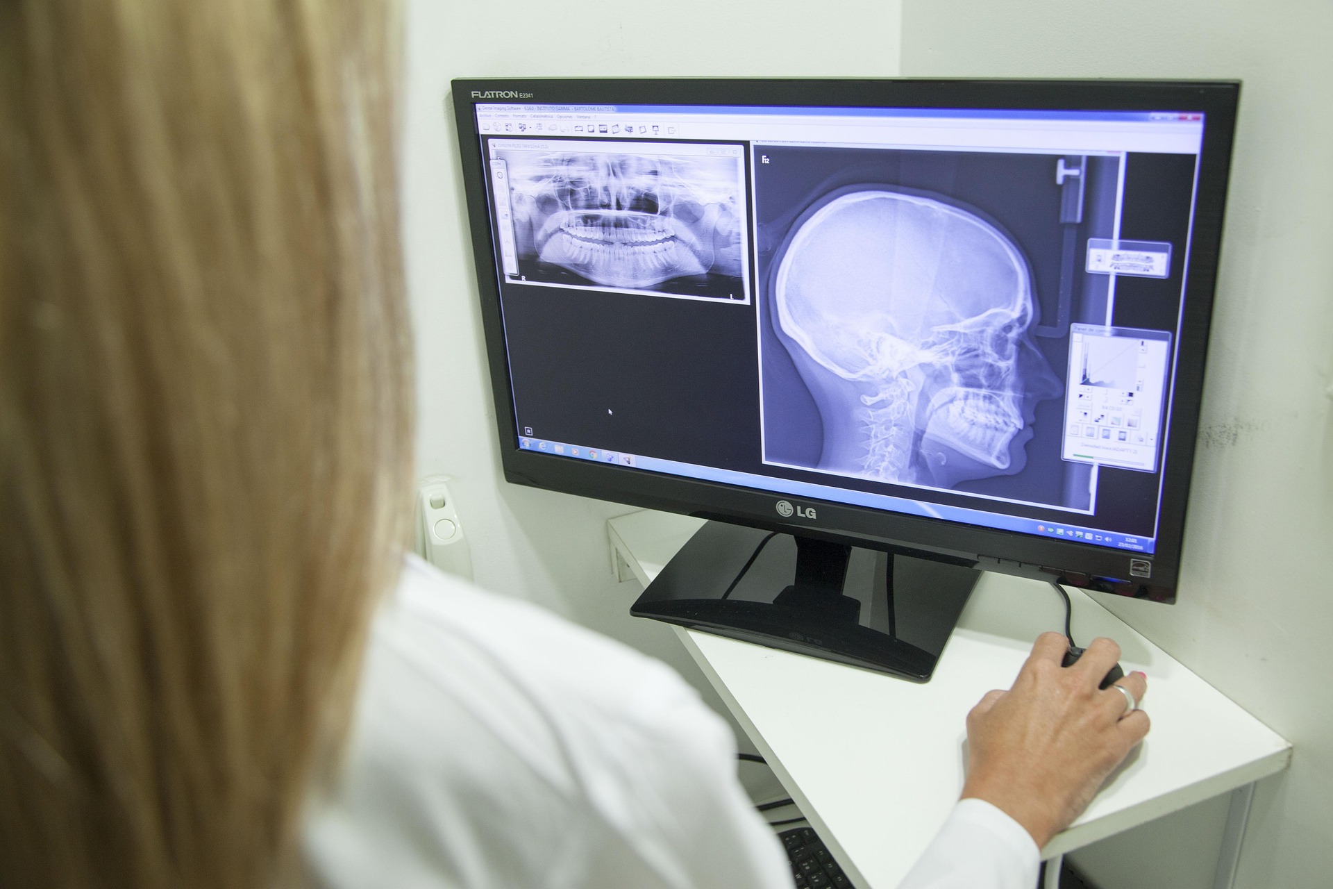
An X-ray is a diagnostic imaging test that uses a small amount of ionising radiation to create images of the inside of the body. Dense structures such as bones absorb more of the X-ray beam, so they appear clearly on the image. Softer tissues appear differently, which allows radiologists and referring doctors to assess certain organs, joints, and internal structures.

X-rays are quick, non-invasive, and widely used because they can provide valuable information in a short space of time. They are especially useful for assessing bones, joints, the chest, spine, and certain abdominal concerns.

While an X-ray does not show every condition in the body, it is often one of the first imaging tests requested because it can confirm, exclude, or guide further investigation. In some cases, your doctor may recommend additional imaging such as ultrasound, MRI, CT, or bone densitometry depending on your symptoms and clinical history.

Why Patients Search for “X-Ray Near Me”

When patients search for X-ray near me, they are usually looking for three things: convenience, trusted reporting, and a radiology practice that can support their doctor’s next steps.

In a busy city like Johannesburg, location matters. Patients may need imaging before returning to work, after visiting a doctor, following an emergency consultation, or as part of ongoing treatment. Access to radiology rooms across areas such as Alberton, Bedford, Sunward Park, and surrounding Johannesburg communities can make the process easier and less stressful.

However, proximity should not be the only consideration. A reliable radiology provider should offer professional imaging, experienced radiologists, clear communication, and proper handling of reports. Diagnostic Radiological Services’ focus is on helping patients receive appropriate imaging in a professional environment, while supporting referring healthcare providers with the information they need.

1. Bone Fractures and Breaks

One of the most common reasons for an X-ray is to check for a fracture. This may happen after a fall, sports injury, car accident, workplace injury, or everyday accident at home.

An X-ray may help diagnose fractures in areas such as:

  • Arms and wrists
  • Hands and fingers
  • Hips and pelvis
  • Legs, knees and ankles
  • Feet and toes
  • Ribs
  • Collarbone
  • Spine

In South Africa, fractures are common across many age groups. Children may need X-rays after playground or school sports injuries. Adults may require imaging after road accidents, gym injuries, or workplace incidents. Older patients may need X-rays after a fall, particularly if osteoporosis or reduced bone strength is a concern.

X-rays can help determine the location, type, and severity of a fracture. This information helps your doctor decide whether you may need a cast, brace, splint, further imaging, specialist referral, or surgical assessment.

2. Joint Dislocations and Alignment Problems

A dislocation happens when the bones in a joint move out of their normal position. It may affect the shoulder, elbow, fingers, hip, knee, ankle, or other joints.

An X-ray can help assess whether a joint is properly aligned and whether there is an associated fracture. This is important because a dislocation may cause damage to surrounding bones, ligaments, and soft tissues.

Common signs that may lead a doctor to request an X-ray include:

  • Sudden joint pain after injury
  • Visible deformity
  • Swelling
  • Difficulty moving the joint
  • Weakness or instability
  • Pain after a joint has been repositioned

If soft tissue damage is suspected, your doctor may recommend further imaging, such as ultrasound or MRI, depending on the joint and the injury.

3. Arthritis and Joint Degeneration

X-rays are commonly used to assess arthritis and degenerative joint changes. They can show changes in joint spacing, bone spurs, and other signs that may suggest wear and tear or inflammatory joint disease.

Patients may be referred for an X-ray if they experience:

  • Ongoing joint pain
  • Morning stiffness
  • Reduced range of motion
  • Swelling around a joint
  • Pain that worsens with activity
  • Chronic knee, hip, hand, shoulder, or spine discomfort

Many patients delay imaging until pain begins affecting daily life. However, early investigation can help your doctor understand whether symptoms are related to arthritis, injury, alignment issues or another condition.

An X-ray does not replace a full medical assessment, but it can provide useful structural information. Your doctor may combine the X-ray findings with your symptoms, blood tests, physical examination, and medical history.

4. Chest Infections and Pneumonia

A chest X-ray is often requested when a doctor needs to assess the lungs, airways, heart, and chest structures. It may be used to help investigate symptoms such as:

  • Persistent cough
  • Shortness of breath
  • Chest pain
  • Fever with breathing symptoms
  • Wheezing
  • Unexplained fatigue
  • Suspected pneumonia

Pneumonia is one of the common conditions that may be assessed with a chest X-ray. The image can help your doctor look for signs of infection, inflammation, or fluid in the lungs.

A chest X-ray may also help when symptoms are not improving as expected or when your doctor needs to check whether further treatment or follow-up imaging is required.

5. Rib Injuries and Chest Trauma

Rib injuries can be extremely painful, especially when breathing, coughing, laughing, or moving. After trauma, a doctor may request an X-ray to check for rib fractures or other chest-related concerns.

This may be necessary after:

  • A fall
  • A sports collision
  • A car accident
  • Direct impact to the chest
  • Workplace trauma
  • Severe coughing with sudden rib pain

While some rib injuries are treated conservatively, imaging can help rule out more serious problems, particularly when pain is severe or breathing is affected. If your doctor suspects internal injury, lung complications, or more complex trauma, CT imaging may be recommended.

6. Spine Conditions and Back Pain

Back and neck pain are common complaints, especially among people who sit for long hours, perform physically demanding work, play sport, or have had previous injuries. An X-ray may be used to assess the bones of the spine and identify structural concerns.

X-rays may help evaluate:

  • Spinal alignment
  • Degenerative changes
  • Scoliosis or curvature
  • Compression fractures
  • Injury after trauma
  • Certain causes of chronic back pain

It is important to understand that X-rays mainly show bones. They do not provide the same level of soft tissue detail as MRI, which is often used to assess discs, nerves, and spinal cord-related concerns. Your doctor will decide which imaging test is most appropriate based on your symptoms.

7. Bone Infections and Abnormal Bone Changes

In some cases, an X-ray may help detect changes related to infection, abnormal growths, or other bone conditions. These findings may not always be visible in the earliest stages, but X-rays can still form part of the diagnostic process.

A doctor may request imaging if there is:

  • Persistent bone pain
  • Swelling or redness
  • A wound near bone
  • Fever with localised pain
  • Pain that does not improve
  • Unexplained changes after previous injury

Depending on the findings, further imaging such as MRI, CT, bone scan, or PET-CT may be recommended. Diagnostic Radiological Services’ broader diagnostic imaging services allow referring doctors to request the most suitable imaging pathway for the patient’s condition.

8. Foreign Objects in Soft Tissue

X-rays can help locate certain foreign objects in the body, particularly if they are dense enough to be seen on imaging. This may include metal fragments, glass, or other materials after an injury.

A doctor may request an X-ray when a patient has:

  • A puncture wound
  • Pain after stepping on or handling a sharp object
  • Swelling after injury
  • Concern that a foreign object remains in the tissue
  • A wound that is not healing properly

Not all materials show clearly on X-rays, so ultrasound or other imaging may be needed in some cases. Your referring doctor will guide the next step depending on the injury and suspected material.

9. Dental, Jaw, and Facial Bone Concerns

Certain X-ray techniques are used to assess dental, jaw, and facial structures. Cone Beam CT may also be used when detailed 3D imaging is required for dental, maxillofacial, or ENT-related assessments.

Patients may require imaging for:

  • Dental infections
  • Impacted teeth
  • Jaw pain
  • Facial trauma
  • Dental implant planning
  • Sinus or facial bone assessment
  • Temporomandibular joint concerns

Because dental and facial structures can be complex, your dentist, maxillofacial surgeon, or specialist may recommend a specific type of imaging depending on the clinical question.

10. Bone Density and Osteoporosis Risk

Bone densitometry, also known as a DEXA scan, is a specialised form of X-ray technology used to measure bone mineral density. It helps assess osteoporosis and osteopenia, which are conditions linked to reduced bone strength.

This is especially relevant for:

  • Post-menopausal women
  • Older adults
  • Patients with a history of fractures
  • People on certain long-term medications
  • Patients with medical conditions affecting bone health
  • Individuals with a family history of osteoporosis

In South Africa, osteoporosis can be underdiagnosed until a fracture occurs. Bone densitometry helps doctors assess fracture risk and manage bone health proactively.

11. Abdominal Concerns

An abdominal X-ray may be requested when a doctor needs to assess certain internal structures. While ultrasound, CT, or MRI may be better suited for many abdominal conditions, an X-ray can still be useful in specific situations.

An abdominal X-ray may help investigate:

  • Severe abdominal pain
  • Suspected bowel obstruction
  • Certain swallowed objects
  • Follow-up after specific procedures
  • Some urinary or digestive concerns

Because abdominal symptoms can have many causes, your doctor will decide whether an X-ray is enough or whether another imaging test is more suitable.

12. Follow-Up After Treatment or Surgery

X-rays are often used to monitor healing and treatment progress. For example, your doctor may request follow-up imaging after a fracture to check whether the bone is healing correctly.

Follow-up X-rays may be used after:

  • Casting or bracing
  • Orthopaedic surgery
  • Joint replacement
  • Spinal procedures
  • Chest infections
  • Injury recovery
  • Medical device placement

This type of imaging helps your healthcare provider confirm whether the treatment plan is working or whether changes are needed.

What to Expect During an X-Ray

Most standard X-rays are quick and straightforward. Depending on the body part being examined, you may be asked to stand, sit, or lie down. The radiographer will position you carefully so the correct area can be imaged.

You may be asked to remove jewellery, belts, or clothing with metal parts, as these can interfere with the image. For chest X-rays, you may be asked to take a deep breath and hold it briefly while the image is taken.

The procedure is usually painless, although positioning may be uncomfortable if you are injured. The imaging itself only takes a short time, and you can usually return to normal activities afterwards unless your doctor advises otherwise.

After the examination, a radiologist reviews the images and prepares a report. This report is made available to your referring healthcare provider so they can discuss the findings and next steps with you.

Is an X-Ray Safe?

X-rays use a small amount of ionising radiation. In medical imaging, the aim is always to use the appropriate test for the clinical need while keeping exposure as low as reasonably achievable.

Your doctor will refer you for an X-ray when the expected diagnostic benefit outweighs the risk. It is important to tell the radiographer if you are pregnant, think you may be pregnant, or have any concerns before the examination.

Children may also have X-rays when clinically necessary, but special care is taken to use appropriate imaging techniques for younger patients.

When Should You Search for “X-Ray Near Me”?

You may need to search for X-ray near me if your doctor has given you a referral or if you have been advised to get imaging after an injury or illness. Common reasons include pain, swelling, trauma, ongoing cough, chest symptoms, suspected fracture, or follow-up after treatment.

You should seek urgent medical attention if you have severe pain, chest pain, difficulty breathing, a serious injury, sudden weakness, confusion, or symptoms that feel like an emergency. In these cases, do not delay care while searching online.

For non-emergency referrals, choosing a trusted radiology practice helps ensure that your imaging is performed professionally and reported by qualified radiologists.

Why Choose Diagnostic Radiological Services for X-Rays?

When patients search for X-ray near me, they need a provider that combines accessibility with professional radiology expertise. Diagnostic Radiological Services provides diagnostic imaging support across multiple locations in Johannesburg’s east and south, serving both outpatient and hospital-based patients.

Our services include general X-ray imaging as well as a broader diagnostic offering, allowing doctors to refer patients for additional imaging when required. This is especially valuable when symptoms are complex or when a first-line X-ray suggests the need for further assessment.

Patients and referring doctors choose Diagnostic Radiological Services because we focus on:

  • Professional diagnostic imaging
  • Experienced radiology teams
  • A wide range of imaging services
  • Support for both routine and specialised referrals
  • Clear reporting to assist clinical decision-making
  • Accessible locations for communities in and around Johannesburg

FAQs About X-Rays

What conditions can an X-ray diagnose?

An X-ray can help diagnose or assess conditions such as bone fractures, joint dislocations, arthritis, chest infections, pneumonia, rib injuries, spinal alignment issues, foreign objects, and certain dental or facial bone concerns. Your doctor will decide whether an X-ray is the right test for your symptoms.

Do I need a referral for an X-ray?

In most cases, X-rays are requested by a doctor, dentist, specialist, or other healthcare provider. A referral helps ensure the correct body part is imaged and that the radiologist understands the clinical reason for the examination.

How long does an X-ray take?

A standard X-ray is usually quick. The imaging itself may only take a few minutes, although registration, preparation, and positioning can add extra time. More complex examinations may take longer.

Is an X-ray painful?

The X-ray itself is not painful. However, positioning may be uncomfortable if you have an injury or severe pain. The radiographer will help position you as carefully as possible.

Is an X-ray safe?

X-rays use a small amount of ionising radiation. They are widely used when medically justified. Your healthcare provider will request an X-ray when the expected benefit of the information gained is greater than the potential risk.

Can I have an X-ray if I am pregnant?

You should tell the radiographer if you are pregnant or think you may be pregnant before any X-ray. Your doctor and radiology team will consider the safest and most appropriate imaging option.

What is the difference between an X-ray and a CT scan?

An X-ray creates a flat image of a specific area, while a CT scan uses X-ray technology to create detailed cross-sectional images. CT is often used when more detail is needed, especially for complex injuries, internal organs, or certain medical conditions.

What is the difference between an X-ray and an MRI?

An X-ray is especially useful for bones and chest imaging. MRI uses magnetic fields and radio waves to create detailed images of soft tissues such as muscles, ligaments, discs, nerves, and organs. Your doctor will choose the most appropriate test based on your symptoms.

What should I wear for an X-ray?

Wear comfortable clothing without metal parts if possible. You may be asked to remove jewellery, belts or clothing that could interfere with the image.

Where can I find an X-ray near me in Johannesburg?

If you are searching for X-ray near me in Johannesburg’s eastern or southern areas, Diagnostic Radiological Services offers radiology support across multiple branches, including areas such as Alberton, Bedford and Sunward Park. Contact us to confirm the most suitable location for your referral.

Book Your X-Ray With a Trusted Radiology Team

If your doctor has referred you for an X-ray, choosing the right radiology provider can make the process easier and more reassuring. Diagnostic Radiological Services supports patients with professional imaging, experienced reporting, and access to a wide range of diagnostic services.

Whether you are dealing with an injury, ongoing pain, chest symptoms, a suspected fracture, or a follow-up referral, our team can assist with the imaging your healthcare provider needs.

When you search for  X-ray near me , choose a radiology team that understands the importance of accurate imaging, patient care, and timely support.

Contact Diagnostic Radiological Services to book your X-ray or speak to our team about the most suitable branch for your referral.

Benefits of Minimally Invasive Interventional Treatments

Modern medicine is moving towards treatments that are more precise, less disruptive, and designed around the patient’s comfort and recovery. For many South Africans, this shift has made interventional treatment an important part of healthcare, particularly when a condition can be managed through a minimally invasive, image-guided procedure rather than traditional open surgery.

Diagnostic Radiological Services understands that patients want clear answers, trusted expertise, and treatment options that support their quality of life. Through advanced imaging technology and radiologist-led care, interventional radiology allows us to diagnose and treat certain conditions through small entry points in the skin, often with less trauma to the body.

Whether a patient has been referred for a biopsy, a vascular procedure, tumour ablation, or fibroid treatment, minimally invasive interventional treatment can offer meaningful benefits when clinically appropriate. These procedures are guided by imaging such as X-ray, ultrasound, or CT, allowing the specialist to see inside the body in real time and target the area of concern with accuracy.

This article explains what minimally invasive interventional treatments are, how they work, and why they are becoming increasingly valuable in South African healthcare.

What Is Interventional Treatment?

Interventional treatment refers to a medical procedure performed with the help of imaging guidance. Instead of making a large surgical incision, an interventional radiologist uses imaging technology to guide small instruments, needles, catheters, or treatment devices to a specific area inside the body.

This approach is commonly known as interventional radiology. It combines diagnostic imaging expertise with targeted treatment. The aim is to treat certain medical conditions as precisely as possible while reducing the physical impact on the patient.

Depending on the condition and the referral from the treating doctor, interventional treatment may be used for procedures such as:

  • Image-guided biopsies
  • Tumour ablations
  • Vascular treatments
  • Uterine fibroid embolisation
  • Other specialist procedures where imaging guidance is required

Not every patient or condition is suitable for interventional radiology, which is why assessment and referral are essential. The treating doctor and radiology team consider the patient’s diagnosis, medical history, imaging results, and overall health before determining whether a minimally invasive option is appropriate.

Why Minimally Invasive Treatment Matters

For many patients, the idea of undergoing a procedure can be stressful. Concerns about hospital admission, pain, downtime, scarring, and complications are completely understandable. Minimally invasive interventional treatment helps address some of these concerns by focusing on targeted care.

Instead of opening a large area of the body to reach the treatment site, imaging allows the specialist to navigate internally with precision. This can reduce unnecessary disruption to surrounding tissue and may support a smoother recovery process.

In a busy South African context, where patients often need to return to work, family responsibilities, and daily life as soon as safely possible, this can make a practical difference. While every patient’s recovery is unique, minimally invasive options can often offer a less intensive experience than conventional surgery.

The Role of Imaging in Interventional Treatment

The strength of interventional radiology lies in image guidance. Imaging is not simply used before or after the procedure; it is part of the treatment itself.

At Diagnostic Radiological Services, imaging modalities such as X-ray, ultrasound, and CT play an important role in guiding procedures. These technologies allow the radiologist to identify the correct area, monitor movement inside the body, and deliver treatment with accuracy.

This precision is one of the major reasons interventional treatment is so valuable. The specialist can work with a clear view of the anatomy, helping to reduce guesswork, and improve procedural control.

For patients, this means the procedure is planned and performed with detailed visual information. For referring doctors, it provides confidence that the treatment is supported by radiological expertise and high-quality imaging.

Benefit 1: Smaller Incisions and Less Physical Trauma

One of the clearest benefits of minimally invasive interventional treatment is that it usually requires only a small puncture or entry point rather than a large incision.

This can help reduce trauma to the body. Smaller access points may mean less disruption to skin, muscle, and surrounding tissue. In many cases, this also means fewer visible marks after healing when compared with open surgical procedures.

For patients who feel anxious about surgery, this can make the treatment journey feel more manageable. A smaller physical intervention can also support comfort during the recovery period, although the exact experience depends on the type of procedure performed and the patient’s individual health profile.

Benefit 2: Image-Guided Precision

Precision is central to interventional radiology. Because the procedure is guided by imaging, the radiologist can target the area of concern directly.

This is particularly important in procedures such as biopsies, tumour ablations, and vascular treatments, where accuracy matters. The ability to see the treatment area in real time helps the specialist reach the correct location while avoiding unnecessary interference with nearby structures as far as possible.

For patients, this targeted approach can offer reassurance. For doctors, it provides valuable support in diagnosis, treatment planning and patient management.

Benefit 3: Faster Recovery in Suitable Cases

Many minimally invasive procedures are associated with shorter recovery times when compared with more invasive surgical approaches. Because there is generally less tissue disruption, the body may have less healing to do.

This does not mean every patient will recover at the same speed, and it does not mean there are no risks. Recovery depends on the specific procedure, the patient’s condition, aftercare instructions, and the advice of the treating doctor.

However, when interventional treatment is suitable, patients may benefit from a more efficient return to normal activities. This can be especially important for working adults, caregivers, parents, and patients managing ongoing health responsibilities.

Benefit 4: Reduced Need for Open Surgery

In some cases, interventional treatment may provide an alternative to open surgery. This is one of the reasons it has become such an important field within modern radiology.

For example, uterine fibroid embolisation is a minimally invasive procedure used to treat fibroids. Instead of surgically removing the uterus or fibroids, embolisation works by targeting the blood supply to the fibroids, helping to shrink them over time. This type of treatment may be considered for suitable patients after assessment and referral.

The benefit is not simply that the procedure is less invasive. It is that the patient may have access to another treatment pathway, one that can be discussed with their healthcare provider based on their symptoms, diagnosis, and future health goals.

Benefit 5: Improved Patient Comfort

Patient comfort is an important part of healthcare. While no medical procedure is completely stress-free, minimally invasive approaches are often designed to reduce the intensity of the experience.

Because interventional treatment is targeted and usually involves smaller access points, patients may experience less discomfort than they expect from traditional surgery. Procedures are also carefully planned, with the radiology team explaining preparation, positioning, and aftercare as needed.

Clear communication matters. Patients should feel able to ask questions, understand why the procedure is being recommended and know what to expect before, during, and after treatment.

Benefit 6: Shorter Hospital Stays in Many Cases

Depending on the procedure and the patient’s condition, some minimally invasive treatments may require a shorter stay than traditional surgery. Certain procedures may be performed without an extended hospital admission, while others may still require monitoring.

This can be valuable for patients who want to recover in the comfort of their own homes once it is medically safe to do so. It can also reduce disruption to family and work routines.


The decision is always clinical. A patient should follow the guidance of their doctor and radiology team regarding observation, discharge, and aftercare.

Benefit 7: A Strong Link Between Diagnosis and Treatment

Diagnostic imaging is often the first step in understanding what is happening inside the body. Interventional radiology builds on this by using imaging not only to identify a problem, but also to guide treatment.

This creates a strong connection between diagnosis and care. A radiologist’s understanding of anatomy, imaging interpretation, and procedural guidance becomes central to the patient’s treatment journey.

Diagnostic Radiological Services’ broader diagnostic offering supports this approach. Services such as X-rays, ultrasound, mammography, bone densitometry, MRI, CT, PET-CT, and cone beam CT help provide the imaging foundation that many patients and doctors rely on for informed decision-making.

Benefit 8: Support for Referring Doctors

Interventional radiology is part of a wider healthcare team. Patients are typically referred by a doctor, and the results of imaging or procedures help guide the next steps in care.

After an examination or procedure, images are reviewed by a radiologist and a report is made available through the appropriate channels. This helps the referring doctor interpret findings and advise the patient on treatment, follow-up, or further investigation.

This collaborative model is important. It ensures that interventional treatment is not viewed in isolation, but as part of a patient’s overall medical care.

Benefit 9: Treatment Options for Specific Conditions

Minimally invasive interventional treatments can be used in several areas of healthcare. The exact procedure depends on the patient’s condition and referral.

Fibroid Treatment

Fibroids can cause symptoms such as heavy bleeding, pelvic discomfort, pressure symptoms, and quality-of-life concerns. For suitable patients, uterine fibroid embolisation may offer a minimally invasive treatment option guided by interventional radiologists.

Image-Guided Biopsies

When a doctor needs a tissue sample to support diagnosis, an image-guided biopsy can help target the area of concern. Imaging guidance supports accuracy and can help reduce unnecessary tissue disruption.

Tumour Ablation

Tumour ablation is a specialised interventional procedure that uses targeted treatment to destroy abnormal tissue. It is considered in specific clinical situations and requires careful assessment.

Vascular Treatments

Interventional radiology may also assist with vascular treatments where imaging guidance is needed to access and treat blood vessels. These procedures are highly specialised and depend on the patient’s diagnosis.

Why South African Patients Are Considering Interventional Treatment

South African patients are increasingly informed about their healthcare options. Many want to understand whether there are alternatives to major surgery, especially when they are balancing work, family, and financial responsibilities.

Interventional treatment can be especially appealing because it combines modern imaging with targeted procedural care. It may reduce downtime, support a more comfortable recovery, and provide treatment options for conditions that previously required more invasive procedures.

Access to experienced radiology services also matters. Patients in Johannesburg and surrounding areas benefit from having radiology practices that support both diagnostic imaging and interventional radiology, helping referring doctors and patients access essential services closer to home.

What to Expect Before an Interventional Procedure

Preparation depends on the specific procedure. Patients may need to pre-book, obtain medical aid authorisation, and bring a referral note from their doctor. They may also be asked to provide medical aid details, date of birth, the reason for the study, and the relevant ICD-10 code where required.

Before the procedure, the team may explain:

  • What the procedure involves
  • Which imaging method will be used
  • Whether fasting or medication instructions apply
  • What to wear or bring on the day
  • Whether someone should accompany the patient
  • What aftercare may be required

Patients should always disclose allergies, current medication, pregnancy status, previous reactions to contrast, chronic conditions, and any relevant surgical history.

What Happens After Interventional Treatment?

After the procedure, patients may be monitored for a period, depending on the type of treatment performed. The radiology team or referring doctor will provide instructions about rest, wound care, medication, activity restrictions, and follow-up.

A radiologist reviews the imaging and procedural findings, and the results or report are made available to support the patient’s ongoing care. In many cases, the referring doctor will discuss the findings and next steps with the patient.

Patients should contact their healthcare provider if they experience concerning symptoms after a procedure or if they are unsure about their aftercare instructions.

Is Interventional Treatment Safe?

All medical procedures carry some level of risk. The advantage of minimally invasive interventional treatment is that it is carefully planned and guided by imaging, which helps improve precision and procedural control.

Safety depends on several factors, including:

  • The type of procedure
  • The patient’s medical history
  • The condition being treated
  • The imaging method used
  • The expertise of the clinical team
  • Proper preparation and aftercare

Patients should always discuss risks, benefits, and alternatives with their doctor before proceeding. A clear understanding of the treatment plan helps patients make informed healthcare decisions.

Who May Benefit from Interventional Treatment?

A patient may benefit from interventional treatment if their condition can be managed through an image-guided procedure and their doctor considers them a suitable candidate.

This may include patients who:

  • Need a biopsy for diagnostic confirmation
  • Have fibroids and are exploring minimally invasive treatment options
  • Require a specialised vascular procedure
  • Need targeted tumour treatment in appropriate clinical circumstances
  • Want to understand alternatives to traditional surgery
  • Have been referred by a doctor for interventional radiology assessment

Suitability is always assessed individually. What works for one patient may not be right for another.

Why Choose Diagnostic Radiological Services?

We combine advanced imaging with radiology expertise to support accurate diagnosis and carefully guided procedures. Our team works with referring doctors to help patients access appropriate imaging and interventional care.

Our services are built around accuracy, patient care, and clinical support. With a range of diagnostic imaging services and interventional radiology capabilities, we aim to make the patient journey as clear and reassuring as possible.

We also understand the importance of accessibility. With multiple branches serving areas in the east and south of Johannesburg, patients can access trusted radiology services closer to where they live and work.

Speak to Us About Interventional Treatment Options

If your doctor has recommended an image-guided procedure, or if you would like to understand whether minimally invasive interventional treatment may be suitable for your condition, our team is here to assist.

We encourage patients to speak to their referring doctor and contact us for guidance on bookings, preparation, and the information needed before an appointment. With radiologist-led expertise and advanced imaging support, we are committed to helping patients move forward with confidence.

FAQs About Interventional Treatment

What is interventional treatment?

Interventional treatment is a minimally invasive medical procedure performed with imaging guidance. It allows a radiologist to diagnose or treat certain conditions using tools such as needles or catheters, often through a small entry point in the skin.

Is interventional treatment the same as surgery?

Interventional treatment is not the same as traditional open surgery. It is usually less invasive and uses imaging such as X-ray, ultrasound, or CT to guide the procedure. However, it is still a medical procedure and must be recommended by a qualified healthcare provider.

What conditions can be treated with interventional radiology?

Interventional radiology may be used for image-guided biopsies, tumour ablations, vascular treatments, and fibroid treatment such as uterine fibroid embolisation. Suitability depends on the patient’s diagnosis and medical assessment.

Is interventional treatment painful?

Patient experience varies depending on the procedure. Many minimally invasive treatments are designed to reduce discomfort compared with more invasive surgery. The radiology team will explain what to expect and how discomfort is managed.

How do I know if I am a candidate for interventional treatment?

Your referring doctor will assess your symptoms and medical history, and imaging may be required to determine whether interventional treatment is suitable. The final decision depends on the condition, the treatment goal, and your overall health.

Do I need a referral for interventional treatment?

Yes, patients generally require a referral from a doctor. The referral helps the radiology team understand the reason for the procedure and ensure that the correct imaging or treatment pathway is followed.

How should I prepare for an interventional procedure?

Preparation depends on the procedure. You may need to pre-book, obtain medical aid authorisation, bring your referral note, and follow specific instructions about medication, fasting, or aftercare. The team will guide you on what is required.

What happens after the procedure?

After the procedure, you may be monitored for a period. A radiologist will review the relevant imaging and findings, and your referring doctor will use the results to guide your next steps.

Is interventional treatment available in Johannesburg?

Yes, Diagnostic Radiological Services supports patients across multiple locations in the east and south of Johannesburg, offering a range of diagnostic imaging services and interventional radiology support.

Why is imaging important during interventional treatment?

Imaging allows the radiologist to see inside the body during the procedure. This helps guide instruments accurately to the target area, supporting precision and reducing unnecessary disruption to surrounding tissue.

The Future of Patient-Centred Treatment

The future of healthcare is increasingly precise, personalised, and patient-centred. Interventional radiology reflects this shift by using imaging to guide treatment directly to the area that needs attention.

For many South Africans, minimally invasive interventional treatment offers a valuable combination of accuracy, comfort, and reduced disruption. It can help patients access advanced care while supporting a more efficient recovery journey in suitable cases.

Diagnostic Radiological Services is proud to support this modern approach to radiology. Through advanced imaging, specialist expertise, and compassionate care, we help patients and doctors make informed decisions at important moments in the healthcare journey.

Choosing the Right Diagnostic Services for Your Condition

When something does not feel right with your body, the first step is often a consultation with a doctor. The next step may be a diagnostic imaging examination that helps your healthcare provider understand what is happening beneath the surface. This is where professional diagnostic services play such an important role in modern healthcare.

Diagnostic Radiological Services understands that patients are often referred for imaging during uncertain, stressful or time-sensitive moments. You may be dealing with pain, a suspected injury, a new symptom, a screening concern or a follow-up after previous treatment. Whatever the reason, the right scan can help your doctor make better-informed decisions about your care.

Diagnostic imaging is not a one-size-fits-all service.

  • An X-ray may be ideal for a suspected fracture.
  • An ultrasound may be suitable for soft-tissue, abdominal, pelvic or pregnancy-related imaging.
  • MRI may be recommended for detailed soft-tissue, joint, spine or neurological assessment.
  • CT may be used when fast, detailed cross-sectional images are needed.
  • Mammography supports breast screening and breast health assessment
  • Bone densitometry helps evaluate bone strength and fracture risk.

Choosing the right diagnostic services begins with understanding what each modality does, why your doctor may request it, and how to prepare for your appointment.

Understanding the Role of Diagnostic Services in Healthcare

Diagnostic services help doctors investigate symptoms, confirm or rule out conditions, monitor known medical concerns and plan treatment. In radiology, these services use imaging technology to create detailed views of the inside of the body.

For patients, imaging can provide clarity and help doctors to provide evidence. In treatment teams, it can guide the next step.

In South Africa, where patients may move between GPs, specialists, emergency care, private healthcare providers and medical aid processes, reliable diagnostic services help create a clearer clinical picture. A scan report can support referrals, treatment planning, surgery preparation, specialist consultations and follow-up care.

Our role is to provide accurate imaging, professional radiological interpretation and a patient-centred experience across a wide range of diagnostic needs.

Why the “Right” Diagnostic Service Matters

The best diagnostic service is the one that is appropriate for your symptoms, clinical history and doctor’s request. Different imaging tests show different parts of the body in different ways.

For example, bones and chest structures may be assessed effectively with X-ray imaging. Soft tissues, tendons, organs and blood flow may be better suited to ultrasound. Complex internal structures may require CT or MRI. Breast health may require mammography, sometimes combined with ultrasound. Bone health may require bone densitometry.

Choosing the correct scan matters because it can:

  • Improve diagnostic accuracy
  • Help avoid unnecessary tests
  • Support faster medical decision-making
  • Assist with treatment planning
  • Reduce delays in care
  • Provide clearer follow-up information
  • Help doctors compare current and previous imaging

This is why your referring doctor’s clinical request is so important. The referral helps the radiology team understand what needs to be investigated and which examination is most suitable.

Common Types of Diagnostic Services and When They Are Used

X-Ray Imaging

X-rays are among the most commonly used diagnostic services. They are quick, widely used and helpful for assessing bones, joints, the chest and certain abdominal concerns.

Your doctor may request an X-ray for:

  • Suspected fractures
  • Joint injuries
  • Chest symptoms
  • Spinal alignment concerns
  • Arthritis-related changes
  • Follow-up after certain treatments or injuries

X-rays are often the first imaging step because they are efficient and effective for many common conditions. They can also help determine whether more advanced imaging, such as CT or MRI, may be needed.

Ultrasound

Ultrasound uses sound waves to create images of internal structures. It does not use ionising radiation, which makes it suitable for many different patients and clinical situations.

Ultrasound may be used to assess:

  • Abdominal organs
  • Pelvic structures
  • Pregnancy-related concerns
  • Soft-tissue lumps or swelling
  • Tendons and muscles
  • Blood flow in certain vessels
  • Breast concerns alongside mammography

Ultrasound is dynamic, meaning the radiologist or sonographer can assess certain structures in real time. This makes it particularly useful for soft tissue, fluid collections and movement-related concerns.

Mammography

Mammography is a specialised breast imaging service used for breast screening and diagnostic breast assessment. It can detect changes in breast tissue that may not be felt during a physical examination.

Your doctor may recommend mammography for:

  • Routine breast screening
  • A new breast lump
  • Breast pain or changes
  • Nipple changes or discharge
  • Follow-up after previous breast imaging
  • Family history or increased breast cancer risk

In many cases, mammography may be combined with breast ultrasound for a more complete assessment, depending on age, breast density, symptoms and clinical findings.

Breast health can feel deeply personal, and we understand that patients may feel nervous before a mammogram. Our team aims to make the process as respectful, efficient and comfortable as possible.

Bone Densitometry

Bone densitometry, often called a bone density scan, helps assess bone strength. It is commonly used to evaluate osteoporosis risk or monitor bone health over time.

A bone density scan may be recommended if you:

  • Have had a low-impact fracture
  • Are post-menopausal
  • Have risk factors for osteoporosis
  • Use certain long-term medications
  • Have a medical condition affecting bone health
  • Need follow-up monitoring for existing bone density concerns

This type of diagnostic service is especially valuable because bone loss often develops silently. A scan can help your doctor identify risk early and recommend lifestyle, medical or treatment interventions where appropriate.

CT Scans

Computed tomography, uses X-rays and computer processing to create detailed cross-sectional images of the body. CT is often used when doctors need fast, detailed information.

A CT scan may be requested for:

  • Trauma or injury assessment
  • Chest, abdominal or pelvic concerns
  • Complex bone injuries
  • Certain vascular assessments
  • Cancer diagnosis, staging or follow-up
  • Infection or inflammatory conditions
  • Emergency or urgent diagnostic questions

Some CT scans require contrast, depending on what your doctor needs to investigate. Contrast helps highlight blood vessels, organs or specific tissues. If contrast is required, the radiology team will guide you on preparation and safety considerations.

MRI Scans

MRI, or magnetic resonance imaging, uses a powerful magnetic field and radio waves to create detailed images. It is particularly useful for soft tissue, joints, the spine, brain and certain internal structures.

MRI may be recommended for:

  • Back or neck pain
  • Disc and nerve concerns
  • Brain or neurological symptoms
  • Joint injuries
  • Sports injuries
  • Soft-tissue abnormalities
  • Pelvic or abdominal assessment
  • Follow-up after surgery or treatment

MRI scans usually take longer than X-rays or CT scans. Some patients may also need preparation if they have implants, pacemakers or metal fragments in the body. Because MRI uses a strong magnetic field, safety screening is an important part of the process.

PET-CT

PET-CT combines metabolic and anatomical imaging. It is often used in more specialised diagnostic pathways, particularly in oncology and certain complex medical investigations.

Doctors may request PET-CT to help with:

  • Cancer staging
  • Treatment response assessment
  • Detection of recurrence
  • Certain infection or inflammatory investigations
  • Specialist-directed diagnostic planning

PET-CT is usually requested by a specialist and forms part of a broader clinical assessment.

Cone Beam CT

Cone Beam CT provides specialised three-dimensional imaging, often used in dental, maxillofacial and certain ENT-related assessments. It can provide detailed views of teeth, jaws, sinuses and facial structures.

This service may be useful for:

  • Dental implant planning
  • Jaw and facial bone assessment
  • Impacted teeth
  • Sinus-related evaluation
  • Specialist dental or maxillofacial referrals

Because Cone Beam CT is highly detailed for specific anatomical areas, it can be valuable when standard dental X-rays are not enough.

How Your Symptoms May Influence the Right Imaging Choice

Patients often search for diagnostic services because they know something is wrong but are unsure which scan they need. The correct imaging choice depends on your symptoms, medical history and doctor’s clinical examination.

For example, sudden ankle pain after a fall may begin with an X-ray. Ongoing shoulder pain with suspected tendon involvement may require ultrasound or MRI. Abdominal pain may be assessed with ultrasound or CT, depending on the clinical concern. A breast lump may require mammography and ultrasound. Unexplained weight loss, cancer follow-up or complex symptoms may require more advanced imaging such as CT, MRI or PET-CT.

The most important step is to follow your doctor’s referral. If the requested examination needs adjustment based on your symptoms or safety considerations, our team can help guide the process within the correct clinical framework.

What to Consider When Choosing Diagnostic Services in South Africa

Choosing diagnostic services is not only about the type of scan. It is also about quality, accessibility, reporting, patient care and coordination with your referring doctor.

Range of Imaging Services

A radiology practice with a broad range of services can support different diagnostic needs in one network. This is helpful when your doctor needs more than one type of imaging or when your condition requires a step-by-step diagnostic approach.

Diagnostic Radiological Services’s offering includes X-ray, ultrasound, mammography, bone densitometry, CT, MRI, PET-CT and Cone Beam CT, allowing us to support a wide variety of patient and doctor requirements.

Experienced Radiologists

The scan itself is only one part of the process. The interpretation is just as important. A radiologist reviews the images and provides a report that helps your referring doctor understand the findings.

This reporting process is central to quality diagnostic services. It turns imaging into clinical information that can support diagnosis, treatment and follow-up.

Convenient Locations

Accessibility matters, especially when you are unwell, recovering from an injury or managing a busy schedule. Having diagnostic services available across multiple locations in the east and south of Johannesburg makes it easier for patients to access imaging closer to home, work or their referring healthcare provider.

Clear Booking Guidance

Some examinations can be performed on a walk-in basis, while others require pre-booking. Services such as MRI, CT, mammography and bone densitometry often need appointments because they may require preparation, specialised equipment time or specific clinical information.

Booking correctly helps reduce delays and ensures that the right team, equipment and preparation instructions are in place.

Patient Preparation Support

Certain scans require preparation. You may need to fast, drink water, bring previous imaging, avoid certain products before a mammogram, complete a safety questionnaire or provide blood test results if contrast is required.

Good preparation improves image quality, supports safety and helps the appointment run smoothly.

Medical Aid and Administrative Support

In South Africa, many patients need to consider medical aid authorisation, benefit limits, referral requirements and co-payments. Administrative guidance can help patients understand what may be needed before the appointment.

While each medical aid and plan is different, it is always wise to confirm authorisation requirements before advanced imaging such as MRI, CT or PET-CT.

Preparing for Your Diagnostic Imaging Appointment

Preparation depends on the type of diagnostic service you are having. When booking, you should ask whether there are any specific instructions for your examination.

In general, patients should bring:

  • Your doctor’s referral letter
  • Your medical aid details, if applicable
  • Your ID or relevant patient information
  • Previous imaging and reports, if available
  • A list of allergies, especially if contrast may be used
  • Information about implants, devices or previous surgery
  • Any relevant blood results, if requested

For MRI, you will usually complete a safety questionnaire  and  for CT with contrast, additional screening may be required. Mammography, you may be advised not to apply deodorant, lotion or powder on the day of the exam. For ultrasound, you may need a full bladder or fasting, depending on the area being examined.

Preparation may sound like admin, but it makes a real difference. It helps the team produce the best possible images and reduces the chance of needing repeat imaging.

What Happens After Your Scan?

After your examination, the images are reviewed by a radiologist. The radiologist prepares a report for your referring doctor, who will explain the findings in the context of your symptoms, examination and medical history.

It is important to remember that imaging results are only one part of your healthcare picture. Your doctor remains the best person to explain what the findings mean for your condition and what should happen next.

Depending on the results, your doctor may recommend treatment, further tests, specialist referral, follow-up imaging or reassurance if no concerning findings are present.

Diagnostic Services for Preventive Health and Early Detection

Diagnostic services are not only used when something is wrong. They also play a role in preventive healthcare and early detection.

Mammography can support breast cancer screening. Bone densitometry can identify reduced bone density before a serious fracture occurs. Follow-up scans can monitor known conditions. Imaging can also help doctors detect changes earlier, when treatment options may be more effective.

For South African patients, preventive imaging can be especially useful when there is a family history, age-related risk, occupational risk, lifestyle-related risk or a previous medical concern that needs monitoring.

The goal is not to scan unnecessarily. The goal is to use the right diagnostic services at the right time for the right clinical reason.

Why Patients and Doctors Choose Diagnostic Radiological Services

Diagnostic Radiological Services supports patients and referring doctors with a comprehensive range of diagnostic imaging services across multiple branches. Our focus is on accurate imaging, professional reporting and a patient experience that respects the stress and uncertainty that often comes with medical investigations.

We understand that every patient arrives with a different story. Some need urgent clarity after an injury and are managing ongoing symptoms that needs attending routine screening or preparing for specialist treatment. Others are anxious because they do not yet know what their results may show.

Our responsibility is to provide reliable diagnostic services that help move the healthcare journey forward.

Choosing Diagnostic Services With Confidence

When choosing diagnostic services, start with your doctor’s referral and ask the right questions:

  • What condition or concern is being investigated?
  • Which scan has been requested and why?
  • Do I need to book in advance?
  • Is any preparation required?
  • Should I bring previous scans or reports?
  • Will my medical aid require authorisation?
  • When and how will my doctor receive the report?

These questions help you arrive prepared and informed. They also help your healthcare team provide the best possible support.

FAQs About Diagnostic Services

What are diagnostic services?

Diagnostic services are medical tests and procedures used to help doctors identify, confirm or monitor health conditions. In radiology, diagnostic services include imaging examinations such as X-rays, ultrasound, mammography, bone density scans, CT, MRI, PET-CT and Cone Beam CT.

Do I need a doctor’s referral for diagnostic imaging?

In most cases, yes. A referral helps ensure that the correct examination is performed for your symptoms or condition. It also gives the radiologist important clinical information for accurate reporting.

Which diagnostic service is best for my condition?

The best diagnostic service depends on your symptoms, medical history and your doctor’s clinical assessment. X-rays, ultrasound, MRI, CT, mammography and other imaging services each have different strengths. Your doctor will request the examination most appropriate for your condition.

Do I need to book an appointment?

Some diagnostic services may require pre-booking, especially examinations such as MRI, CT, mammography and bone densitometry. It is best to confirm booking requirements before arriving.

Are diagnostic services painful?

Most imaging examinations are not painful. Some may involve mild discomfort, such as positioning during an X-ray, breast compression during mammography or lying still during MRI. The team will guide you through the process and help you feel as comfortable as possible.

What should I bring to my appointment?

Bring your referral letter, ID, medical aid information if applicable, previous imaging or reports, and any relevant medical information. If your scan requires contrast or special preparation, you may be asked to bring additional information.

How long does it take to get results?

After the examination, a radiologist reviews the images and prepares a report. The report is then made available to your referring doctor or according to the practice’s results process. Your doctor will explain the findings and next steps.

Is MRI safer than CT?

MRI and CT are different imaging tools used for different reasons. MRI does not use ionising radiation, while CT uses X-rays to create detailed cross-sectional images. The safest and most appropriate choice depends on your medical condition, urgency, body area being examined and doctor’s request.

What is the difference between screening and diagnostic imaging?

Screening imaging looks for early signs of disease before symptoms appear, such as routine mammography. Diagnostic imaging investigates a specific symptom, abnormal finding or known medical concern.

Can I choose where to have my diagnostic services done?

Yes, patients often have a choice, depending on doctor referral requirements, medical aid rules, location and service availability. Choosing a radiology practice with comprehensive services, experienced radiologists and convenient branches can make the process easier.

Book Professional Diagnostic Services With Diagnostic Radiological Services

When your health depends on clear answers, choosing the right diagnostic services matters. Diagnostic Radiological Services offers a comprehensive range of imaging solutions, including X-rays, ultrasound, mammography, bone densitometry, MRI, CT, PET-CT and Cone Beam CT.

Whether you have been referred for screening, injury assessment, specialist investigation or follow-up imaging, we are here to support you with professional service, advanced imaging options and radiologist-led reporting.

Contact us to book your appointment, confirm preparation requirements or find the branch best suited to your examination.

Advances in Modern Medical Imaging Technology: How Diagnostic Radiology is Transforming Healthcare

Medical imaging has experienced remarkable innovation in recent decades. What once began with simple X-rays has evolved into a sophisticated suite of technologies that allow clinicians to see inside the body with unprecedented clarity, speed, and precision. From early detection of disease to personalised treatment planning and minimally invasive interventions, modern imaging is central to improved patient outcomes.

This article explores the technological breakthroughs shaping modern medical imaging, their clinical applications, and how they are revolutionising healthcare delivery. Whether you are a patient, caregiver, or healthcare professional, understanding these advances helps demystify how imaging guides diagnosis and treatment in today’s medical landscape.

Diagnostic Radiological Services provides a wide range of imaging solutions supported by state-of-the-art equipment and specialist expertise, from basic radiographs to advanced MRI, CT, and functional imaging.

What Is Modern Medical Imaging?

Modern medical imaging refers to advanced diagnostic technologies used to visualise internal structures and physiological processes in the body. These technologies include traditional methods like X-rays and ultrasound, as well as more advanced modalities such as CT (computed tomography), MRI (magnetic resonance imaging), PET (positron emission tomography), and hybrid imaging.

What sets modern medical imaging apart is not just the ability to see inside the body; it is the level of detail, speed of acquisition, and integration with digital tools such as artificial intelligence (AI), 3D reconstruction, and real-time functional imaging.

Key Advances in Medical Imaging Technology

High-Resolution and Low-Dose CT Scanning

Computed tomography (CT) has evolved from simple cross-sectional imaging to advanced scanners capable of capturing high-resolution images of complex anatomy in seconds. Current CT systems offer:

  • Ultra-high-resolution imaging for detailed views of small structures
  • Low-dose protocols that maintain image quality while minimising radiation exposure
  • Dual-energy and spectral CT that differentiate tissue types and enhance contrast
  • Cardiac CT angiography for assessing coronary vessels non-invasively

These advancements make CT indispensable in trauma care, cancer staging, vascular imaging, and cardiopulmonary assessment.

Magnetic Resonance Imaging (MRI) with Faster Sequences and Higher Field Strength

MRI is a radiation-free imaging modality that uses magnetic fields and radio waves to produce detailed soft-tissue images. Progress in MRI technology includes:

  • Faster acquisition sequences that reduce scan time and improve patient comfort
  • High-field MRI (3T and above) for enhanced signal and detailed tissue contrast
  • Functional MRI (fMRI) that maps brain activity based on blood flow
  • Diffusion-weighted imaging (DWI) for early stroke and tumour evaluation

These innovations allow clinicians to differentiate pathology with greater confidence, especially in neurological, musculoskeletal, and oncologic cases.

Artificial Intelligence and Machine Learning Integration

AI and machine learning algorithms are now embedded into imaging workflows to assist radiologists in:

  • Detecting subtle abnormalities
  • Prioritising critical cases for faster review
  • Enhancing image quality and noise reduction
  • Quantifying changes over time for therapy monitoring

AI doesn’t replace human expertise; it augments it, improving accuracy, consistency, and efficiency while reducing diagnostic errors.

Hybrid Imaging: PET/CT and PET/MRI

Hybrid imaging combines the strengths of different modalities into a single scan, offering both anatomical and metabolic information. Examples include:

  • PET/CT: Combines metabolic data from PET with detailed anatomy from CT, commonly used in cancer staging and monitoring
  • PET/MRI: Offers superior soft-tissue contrast alongside metabolic imaging, ideal for neurological and oncologic applications

These hybrid systems help clinicians identify disease earlier, assess treatment response, and personalise care plans.

3D Imaging and Advanced Reconstruction

Modern medical imaging systems can produce three-dimensional (3D) reconstructions that allow clinicians to:

  • Visualise anatomy from any angle
  • Plan surgical or interventional procedures
  • Guide minimally invasive treatments with precision
  • Communicate findings more clearly to patients

3D imaging enhances understanding of complex anatomical relationships, improving both diagnostic confidence and clinical outcomes.

Point-of-Care and Portable Imaging

Small, portable imaging devices are transforming access to diagnostics, particularly in emergency settings, remote areas, and point-of-care environments. Portable ultrasound, handheld X-ray units, and compact CT systems make it possible to bring imaging to the patient, not just the other way around.

Clinical Applications of Advanced Imaging

Modern medical imaging technologies accelerate diagnosis, personalise therapy, and minimise risk. Some key clinical applications include:

  • Emergency Medicine: Rapid trauma assessment with CT, focused ultrasound (FAST), and critical care imaging
  • Oncology: Early detection, tumour staging, treatment response assessment, and follow-up
  • Cardiology: Coronary CT angiography, cardiac MRI for structural assessment
  • Neurology: Stroke imaging, brain tumour evaluation, functional mapping
  • Orthopaedics: High-resolution MRI for joints and soft tissues; CT for complex fractures
  • Women’s Health: Digital mammography, breast MRI for high-risk screening and diagnosis

These applications underscore how modern medical imaging supports comprehensive care pathways across disciplines.

Safety, Comfort, and Patient Experience

While modern medical imaging technology becomes more powerful, patient safety and comfort remain central:

  • Lower radiation doses through optimised protocols
  • Non-ionising imaging options like MRI and ultrasound
  • Faster scan times and open designs reduce anxiety and motion artefacts
  • Sedation and support for claustrophobic or paediatric patients

Imaging facilities now prioritise both diagnostic excellence and the overall experience.

Choosing the Right Imaging Modality

The choice of imaging depends on several clinical factors:

  • The patient’s symptoms and medical history
  • The area of the body being examined
  • The level of detail required
  • Radiation exposure considerations
  • Availability of specialised modalities

Clinicians partner with radiology specialists to determine the most appropriate imaging approach for each situation.

The Radiologist’s Role in Modern Imaging

Radiologists are medical doctors specialising in interpreting imaging studies and integrating them with clinical information. They:

  • Analyse images in detail
  • Generate structured, diagnostic reports
  • Recommend additional imaging or follow-up
  • Collaborate with referring clinicians to guide care decisions

Their expertise ensures imaging contributes meaningfully to diagnosis, treatment, and patient outcomes.

The Future of Medical Imaging

Emerging trends indicate even more transformative advances ahead:

  • Real-time imaging and intervention guidance
  • Radiomics and quantitative imaging biomarkers
  • Deep learning models for pattern recognition
  • Personalised imaging protocols tailored to patient biology

These innovations will continue to enhance precision, speed, and personalised care.

FAQs About Modern Medical Imaging

What are modern medical imaging technologies?

Modern medical imaging includes advanced modalities such as CT, MRI, ultrasound, PET/CT, PET/MRI, 3D imaging, and AI-assisted diagnostics used to view internal structures and functions.

How is AI used in medical imaging?

AI and machine learning help improve image quality, detect subtle findings, prioritise urgent cases, and provide quantitative analysis that supports radiologist interpretation.

Are medical imaging tests safe?

Yes. Imaging is generally safe when performed for clinical reasons. Modalities such as MRI and ultrasound do not use ionising radiation, and X-ray/CT scans use controlled, low doses when medically necessary.

What determines the choice of imaging test?

The specific clinical question, area of interest, required detail, radiation considerations, and patient factors all influence the choice of imaging modality.

How long does it take to get imaging results?

Timing varies; some urgent scans are interpreted immediately, while routine reports may be completed within 24–48 hours. Hybrid or advanced studies may require additional analysis time.

Does medical imaging involve radiation?

Some tests, including X-rays and CT scans, use ionising radiation, while others, like MRI and ultrasound, do not. Your clinician will weigh the benefits and risks before recommending a scan.

Contact Us For Details

As imaging technology continues to evolve, the power of diagnostic radiological services grows, enabling earlier detection, safer care, and better health outcomes. Whether you’re preparing for your first scan or seeking advanced imaging solutions, understanding these advances empowers you to engage meaningfully in your healthcare journey.


Types of Diagnostic Imaging Tests: A Guide to Diagnostic Radiological Services

Diagnostic imaging is one of modern medicine’s most important tools. By creating detailed pictures of the inside of the body, it helps healthcare providers detect injuries, monitor diseases, guide treatment, and make informed decisions about patient care.

Diagnostic radiological services encompass a range of imaging tests, from quick, simple X-rays to advanced 3D imaging such as CT (computed tomography) and MRI (magnetic resonance imaging). Each test has its own strengths, benefits, and clinical uses, and understanding them can help patients know what to expect and why a particular scan may be recommended.

This article explores the key types of diagnostic imaging tests, how they work, what they’re commonly used for, and when they’re most appropriate.

What Are Diagnostic Radiological Services?

Diagnostic radiological services use radiation-based technologies like X-rays, CT scans, and fluoroscopy, as well as non-ionising imaging methods such as MRI and ultrasound to visualise internal structures. These imaging tests play a critical role in diagnosing illnesses, planning treatment, and monitoring progress.

At Diagnostic Radiological Services, highly trained specialists use state-of-the-art equipment to deliver accurate imaging and reliable interpretations in support of clinical care.

Common Types of Diagnostic Imaging Tests

X-Rays

X-rays are the most widely recognised form of diagnostic radiology. They use low doses of ionising radiation to create images of bones and certain tissues.

How they work

When X-ray beams pass through the body, dense structures like bone absorb more radiation and appear white on the image, while softer tissues appear in shades of grey.

Common uses

  • Detecting fractures and dislocations
  • Assessing chest conditions like pneumonia or lung infections
  • Evaluating joint problems and arthritis
  • Guiding certain interventional procedures

When it’s used

X-rays are usually the first line of imaging when bone injury or chest evaluation is needed because they are fast, widely available, and highly effective for these purposes.

Computed Tomography (CT) Scans

CT scans use multiple X-ray beams and computer processing to generate cross-sectional images (slices) of the body.

How they work

As the patient moves slowly through a rotating X-ray beam, multiple images are captured and reconstructed to produce detailed 3D pictures of internal organs, bones, blood vessels, and soft tissues.

Common uses

  • Detecting internal injuries from trauma
  • Diagnosing abdominal and pelvic conditions
  • Evaluating complex bone fractures
  • Detecting tumours and cancers
  • Planning radiation therapy

When it’s used

CT is particularly useful when detailed images are required quickly, for example in emergency settings, cancer detection, or complex internal evaluations.

Magnetic Resonance Imaging (MRI)

MRI uses a powerful magnetic field and radio waves to produce highly detailed images, particularly of soft tissues.

How it works

Instead of ionising radiation, MRI relies on magnetic properties of hydrogen atoms in the body. Radiofrequency pulses and computer algorithms convert signals into detailed images.

Common uses

  • Assessing brain and spinal cord disorders
  • Evaluating joints, ligaments, and cartilage
  • Detecting tumours and soft tissue abnormalities
  • Imaging heart and vascular structures
  • Diagnosing liver and abdominal conditions

When it’s used

MRI is ideal when high-contrast images of soft tissues are needed for neurological, musculoskeletal, and cardiovascular assessments.

Ultrasound

Ultrasound uses high-frequency sound waves to produce real-time images of organs and structures.

How it works

A handheld transducer emits sound waves into the body, which bounce back from internal structures. These echoes are interpreted by a computer to create images.

Common uses

  • Monitoring pregnancy and foetal development
  • Diagnosing gallstones, kidney stones, and abdominal issues
  • Evaluating blood flow using Doppler ultrasound
  • Guiding biopsies and fluid drainage

When it’s used

Ultrasound is safe, non-invasive, and radiation-free, making it ideal for obstetric imaging, vascular studies, and real-time guided procedures.

Mammography

Mammography is a specialised form of X-ray imaging focused on breast tissue.

How it works

Low-dose X-rays capture high-resolution images of the breast, allowing radiologists to detect abnormalities such as microcalcifications and masses.

Common uses

  • Breast cancer screening and early detection
  • Investigating breast lumps or symptoms
  • Monitoring patients with a history of breast lesions

When it’s used

Regular mammograms are recommended for women in age-based screening programmes and for individuals at higher risk of breast cancer.

Choosing the Right Imaging Test

The choice of imaging depends on several factors:

  • The type of symptoms
  • The area of the body being examined
  • The level of detail needed
  • Whether radiation exposure is acceptable
  • Patient age, pregnancy status, or pre-existing conditions

Your healthcare provider will recommend the most appropriate diagnostic radiological services based on clinical findings and imaging requirements.

Preparing for Diagnostic Imaging

Preparation varies by test:

  • X-rays often require no preparation
  • CT scans might require fasting or contrast agents
  • MRI requires screening for metallic implants and possible fasting
  • Ultrasound preparation depends on the area scanned (e.g., a full bladder for pelvic ultrasound)
  • Mammography may require avoiding deodorants or lotions before the test

Your radiology team will provide specific instructions to ensure the best possible imaging results.

How Radiologists Interpret Imaging

Once imaging is complete, a specialist radiologist reviews the images for signs of abnormalities, disease, or injury. A detailed report is then shared with the referring clinician to guide diagnosis and treatment planning.

Quality imaging and expert interpretation are both essential components of effective diagnostic radiological services.

The Role of Diagnostic Imaging in Healthcare

Diagnostic imaging helps healthcare providers:

  • Detect conditions early
  • Plan surgical or interventional procedures
  • Monitor disease progression or treatment response
  • Reduce unnecessary exploratory surgery
  • Improve patient outcomes with accurate, data-driven decisions

From emergency diagnostics to routine screening, imaging plays a central role in modern medicine.

FAQs About Diagnostic Radiological Services

What are diagnostic radiological services?

Diagnostic radiological services involve medical imaging tests like X-rays, CT scans, MRI, ultrasound, and mammography that help diagnose injuries, diseases, and abnormalities inside the body.

How do I know which imaging test I need?

Your doctor determines the best imaging test based on your symptoms, medical history, and the specific area of concern. Each imaging type has strengths for different clinical situations.

Are imaging tests safe?

Yes, most imaging tests are safe. Tests like MRI and ultrasound don’t use ionising radiation. X-rays and CT scans use controlled, low doses, and are performed only when clinically necessary.

Do I need preparation for my imaging test?

Some tests require specific preparation, such as fasting or avoiding certain products. Your radiology provider will give clear instructions based on the test you are having.

How long does it take to get results?

The time varies, but many imaging reports are completed within 24–48 hours. Urgent cases are prioritised and reported faster when needed.

Will my health insurance cover diagnostic imaging?

Coverage varies by plan and policy. It’s best to check with your insurer to understand your benefits for specific imaging services.

Contact Us Today For Details

Diagnostic imaging continues to transform patient care with precise, detailed insight into the body’s internal structures. By understanding the types of Diagnostic Radiological Services available and when they’re used, you can be better informed and more confident in your healthcare journey.


BREAST CANCER RUN 2025